 by Madison Dental Art Review Board
Nearly one in four adults has experienced a cracked or broken tooth, yet when that fracture falls at or below the gum line, the stakes rise sharply. A tooth broken at the gum line is not simply a cosmetic problem. It is a dental emergency that involves the gum tissue, the surrounding bone, and often the nerve inside the root. Comprehending your options for removing a broken tooth at the gum line, or saving it, can mean the difference between a straightforward recovery and months of complicated treatment.
At Madison Dental Art in Forest Hills, Queens, our team has been guiding New York families through exactly these decisions since 1994. This guide explains what happens when a tooth fractures at the gum line, how dentists evaluate the damage, and what modern treatment looks like.
Expert Endodontic Care at Madison Dental Art.
Table of Contents
Key Takeaways
- A tooth broken at the gum line is classified as a crown-root fracture and almost always requires professional evaluation within 24 hours.
- Extraction is not always the only option, several tooth-preserving techniques can save the root and allow a crown to be placed.
- The right treatment depends on how deep the fracture goes, the health of the surrounding bone, and whether infection is present.
- Delaying treatment increases the risk of infection, bone loss, and more complex surgery.
- Replacing a lost tooth promptly protects the neighboring teeth and preserves jaw bone volume.
What Does “Broken at the Gum Line” Actually Mean?
When dentists refer to a tooth broken at the gum line, they use the clinical term crown-root fracture. This means the break extends from the visible crown of the tooth downward through the enamel and dentin, crossing the gum margin and reaching into the root below. In some cases, the fracture line sits just at the edge of the gum tissue. In more severe cases, it extends several millimeters below the bone crest.
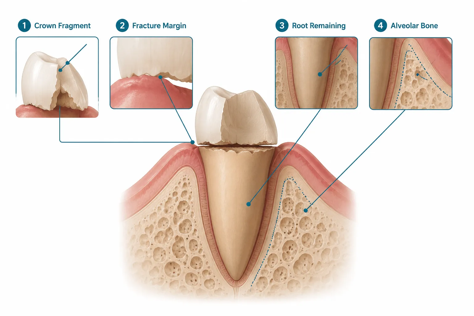
This type of fracture is different from a simple chipped tooth. Because the break involves the area where the gum attaches to the tooth, a zone dentists call the biologic width, treatment becomes considerably more complex. The biologic width is roughly 2 millimeters of tissue that must remain intact for the gum to stay healthy around any restoration. When a fracture invades this space, the dentist cannot simply place a filling or crown without first addressing the gum and bone architecture.
Common Causes
- Biting down on hard foods (ice, hard candy, unpopped popcorn kernels)
- Untreated decay that weakens the tooth from the inside
- Trauma, a fall, sports injury, or accident
- A large existing filling that leaves thin walls of tooth structure
- Grinding and clenching (bruxism) over many years
If you are dealing with a cracked tooth in Forest Hills, Queens, it is worth knowing that not every crack is the same. A crack that stops above the gum line is far easier to manage than one that extends below it.
Diagnosing the Fracture: What to Expect at Your Appointment
Before any treatment plan can be made, the dentist needs a clear picture of exactly where the fracture line sits. This requires more than a visual check.
Diagnostic tools used at Madison Dental Art include:
- Digital X-rays, reveal the depth of the fracture relative to the bone level
- 3D Panoramic Scan, provides a full three-dimensional view of the root, surrounding bone, and adjacent teeth, which is especially useful when the fracture is suspected to be deep
- Clinical probing, the dentist gently probes around the tooth to map the fracture margin and assess bone support
The American Association of Endodontists updated its guidelines for traumatic dental injuries in 2026, emphasizing that timely, accurate diagnosis is the foundation of successful treatment. The updated protocols stress that crown-root fractures should be assessed with both imaging and hands-on clinical evaluation before any treatment decision is made.
If the tooth is painful, you may also want to review how to describe tooth pain to a dentist so your provider can document your symptoms accurately, this helps guide the diagnosis.
Warning Signs That Demand Immediate Attention
| Symptom | What It May Indicate |
|---|---|
| Persistent throbbing pain | Nerve involvement or early infection |
| Swelling in the gum or jaw | Abscess forming below the fracture |
| Visible fragment loosely attached | Unstable crown-root fracture |
| Sensitivity to hot and cold | Pulp (nerve) exposure |
| Bad taste or odor | Bacterial contamination of the fracture site |
If you notice swelling or pus, do not wait. Learn more about signs of tooth infection and contact a dentist the same day. Left untreated, a dental abscess can spread to the jaw and beyond, a risk that is well-documented and serious.
Treatment Options for Removing a Broken Tooth at the Gum Line
This is where the decision-making becomes nuanced. Removing a broken tooth at the gum line does not always mean extracting the entire tooth. In many cases, “removal” refers to removing only the broken coronal fragment while preserving the root. Contemporary dental care in 2026 strongly favors tooth preservation over extraction wherever it is biologically feasible.
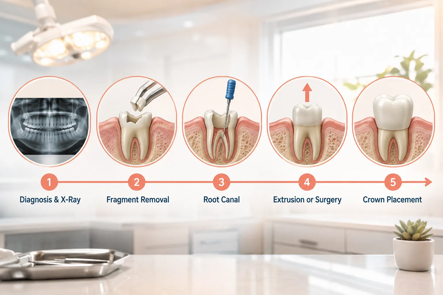
Here are the six main pathways a dental team may consider:
Option 1: Fragment Removal with Supragingival Restoration
If the fracture margin is very close to the gum line, not deeply subgingival, the dentist may remove the loose coronal fragment and restore the tooth with a crown after a minor gingivectomy (trimming of gum tissue). This is the most conservative surgical approach and works best when the fracture does not invade the biologic width significantly.
Option 2: Fragment Removal, Root Canal, and Crown with Gingivectomy or Ostectomy
When the fracture is slightly below the gum but above the bone, the dentist removes the fragment, performs a root canal to protect the nerve, and reshapes the surrounding gum and bone (osseous surgery) to expose enough tooth structure for a crown. This is one of the most common approaches for moderately subgingival fractures.
Option 3: Orthodontic Extrusion (Forced Eruption)
This is a conservative, tooth-saving technique that uses gentle orthodontic force to move the root upward, essentially erupting it through the gum so the fracture margin rises above the tissue level. Research published in 2025 describes applying approximately 50 grams of continuous extrusive force using an elastomeric chain, achieving around 3 millimeters of movement over six weeks, followed by four weeks of retention to allow the bone and gum to remodel. The goal is to create at least 2 millimeters of tooth structure above the gum, called the ferrule, which is essential for a long-lasting crown.
Orthodontic extrusion is particularly valuable for younger patients with healthy roots and strong surrounding bone.
Option 4: Surgical Extrusion
Similar in goal to orthodontic extrusion, surgical extrusion repositions the root upward through a minor surgical procedure rather than over weeks of orthodontic movement. It is faster but requires careful handling to preserve the periodontal ligament fibers that keep the tooth alive.
Option 5: Coronectomy or Root Submergence
In some cases, particularly when a patient is not yet ready for an implant, the dentist may intentionally leave the root beneath the gum and bone. This technique, called root submergence or coronectomy, preserves the alveolar bone volume for a future implant placement. It is a strategic holding option, not a permanent solution.
Option 6: Full Extraction Followed by Replacement
When the fracture extends too deeply into the root, when significant bone loss is present, or when the tooth cannot be restored by any of the above methods, extraction becomes necessary. This is not a failure, it is sometimes the most responsible path forward.
Important: Extraction should not be the automatic first response to a subgingival fracture. Contemporary interdisciplinary literature consistently emphasizes that the natural tooth should be preserved whenever the biologic width and ferrule can be re-established, because doing so protects the surrounding bone, maintains the gum architecture, and preserves the nerve feedback (proprioception) that implants cannot replicate.
If extraction is necessary, knowing what happens if you don’t replace a missing tooth is critical. Bone resorption begins within weeks of losing a tooth, which can affect neighboring teeth and complicate future implant placement.
After Extraction: Replacing the Tooth
If the tooth cannot be saved, the conversation turns immediately to replacement. In 2026, the most reliable long-term options are:
Dental Implant A titanium post is placed into the jaw bone, topped with a custom crown. Implants look, feel, and function like natural teeth and prevent bone loss. Our periodontology specialist, Dr. Robert Weber, a Diplomate of the American Board of Periodontology, leads implant placement at Madison Dental Art. For a detailed overview, see our dental implants procedure timeline.
Dental Bridge A fixed bridge uses the two adjacent teeth as anchors to support an artificial tooth in the gap. It is a faster solution than an implant but requires reshaping the neighboring teeth.
Partial Denture A removable option that replaces one or more missing teeth. Less permanent than an implant or bridge, but a practical solution for some patients.
For patients concerned about recovery, our guide on what to eat after tooth extraction covers the dietary steps that support healing and reduce complications.
Why Prompt Treatment Matters
Every day a broken tooth at the gum line goes untreated, bacteria colonize the exposed fracture site. This accelerates decay, increases the risk of a dental abscess, and can cause the surrounding bone to begin breaking down. Bone loss is particularly damaging because it reduces the foundation available for an implant later, and in some cases, bone grafting becomes necessary before implant placement is even possible.
If you are experiencing persistent tooth pain in Forest Hills or Queens, do not assume it will resolve on its own. A tooth broken at the gum line rarely improves without professional intervention.
How Madison Dental Art Approaches This Treatment
Founded in 1994 by Dr. Alexander Chao, who graduated in the top 3% of his class at NYU College of Dentistry, Madison Dental Art has grown into a full-service practice that handles every stage of a broken tooth case under one roof. From the initial 3D panoramic scan to root canal therapy, periodontal surgery, orthodontic extrusion, and final crown or implant placement, patients do not need to travel across the city to see multiple specialists.
Our team includes:
- Dr. Alexander Chao, general and restorative dentistry, crowns, and overall treatment planning
- Dr. Robert Weber, periodontics and implant surgery, including osseous surgery for subgingival fracture exposure
- Dr. Yutian Zeng, general dentistry with a focus on empathetic, anxiety-aware care
- Dr. Susan Huh, orthodontics, including orthodontic extrusion for tooth preservation
We also offer nitrous oxide sedation for patients who feel anxious about surgical procedures, and our digital imaging suite ensures every treatment decision is based on precise, current data.
Madison Dental Art is located at 112-03 Queens Boulevard, Suite 211, Forest Hills, NY 11375, and is open Monday through Friday, 9:00 AM to 6:00 PM, and Saturday from 8:00 AM to 3:00 PM. Call us at (718) 268-8228 or visit our contact page to schedule an evaluation.
We accept multiple insurance plans, visit our insurance page for details.
FAQs
Can a tooth broken at the gum line always be saved?
Not always, but more often than many patients expect. The main factors are how deep the fracture extends below the gum and bone, the overall health of the root, and whether infection is already present. When the fracture margin can be brought above the gum line through orthodontic or surgical extrusion, and when at least 2 millimeters of healthy tooth structure can be exposed for a crown, the tooth can often be saved.
Is removing a broken tooth at the gum line painful?
With modern local anesthesia and sedation options, the procedure itself should not be painful. Patients may feel pressure during the extraction or surgical extrusion, but sharp pain is not expected. Post-procedure soreness is normal and typically managed with over-the-counter pain relief and the aftercare instructions your dentist provides. For patients with significant dental anxiety, nitrous oxide sedation is available at Madison Dental Art to make the experience more comfortable.
How long does recovery take after a subgingival tooth extraction?
Most patients recover from a straightforward extraction within one to two weeks. Surgical procedures, such as those involving bone reshaping (ostectomy) or orthodontic extrusion, require a longer healing period, often four to eight weeks, before the final restoration can be placed. Bone remodeling after orthodontic extrusion typically takes about four weeks of retention time. Your dentist will provide a personalized timeline based on your specific case.
What is the best replacement option after losing a tooth at the gum line?
For most patients, a dental implant is the gold standard. It is the only replacement option that preserves alveolar bone volume, functions like a natural tooth, and does not require altering adjacent teeth. Nevertheless, the right choice depends on bone availability, overall health, and patient preference. A dental bridge is a viable alternative for patients who want a faster solution, and partial dentures offer a removable option. Your dentist will review all options and help you decide based on your clinical situation and goals.
References
- American Association of Endodontists. (2026). Recommended Guidelines for the Treatment of Traumatic Dental Injuries. https://www.aae.org/specialty/clinical-resources/traumatic-dental-injuries/
- Flores, M. T., et al. (2022). Guidelines for the management of traumatic dental injuries: 3. Injuries in the primary dentition. Dental Traumatology, 38(1), 3-15. https://doi.org/10.1111/edt.12737
- American Academy of Periodontology. (2023). Periodontal disease and systemic health. https://www.perio.org/consumer/gum-disease-and-other-diseases
- National Institute of Dental and Craniofacial Research. (2023). Dental emergencies. U.S. Department of Health and Human Services. https://www.nidcr.nih.gov/health-info/dental-emergencies
- Diangelis, A. J., et al. (2022). International Association of Dental Traumatology guidelines for the management of traumatic dental injuries: 1. Fractures and luxations of permanent teeth. Dental Traumatology, 38(1), 16-29. https://doi.org/10.1111/edt.12740
Madison Dental Art
Content written and reviewed by
Madison Dental Art Review Board
Madison Dental Art : Experienced dentists and specialists dedicated to providing accurate, trusted, and patient-focused oral health information.
Important information
The content of this article is for strictly educational and informational purposes. It does not replace in-person professional medical diagnosis, advice, or treatment.


